Barefoot Photo Gallery
Case 1
Zeik had just had an acute laminitis attack, probably triggered by the frozen grass, and some possible Cushings involvement, when I first saw him in January ’10. His front hooves looked as though he may well have been suffering from chronic low grade laminitis (undiagnosed) for at least several months. His owner said he’d always been a bit ‘grumpy and reluctant’ when ridden. The vet had been involved in this last, more acute attack.
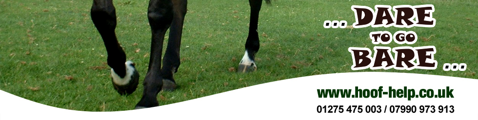
February '10
A tiny mark (a blackish line, less than 5mm long) on his sole, when
gently scrapedwith a hoof pick, gave way to a sub-solar haematoma,
with a possible route into live tissue.
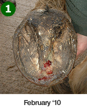
 March
'10
March
'10
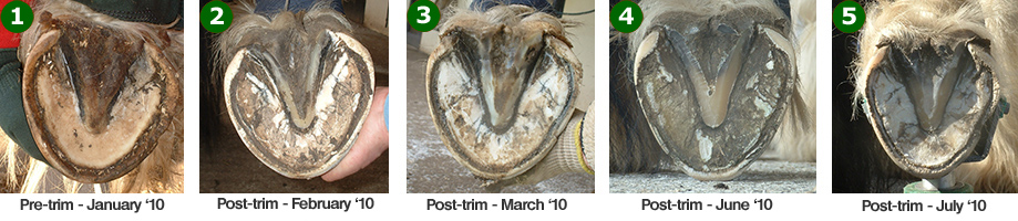
His sole had a ‘hollow’ ring to it when tapped, and began to peel away (with
his frog). He had grown three layers of ‘hung’ sole, showing he had been
trying to heal the damage himself for quite a while. You can see his solar
corium has prolapsed through some of the layers of sole. Also notice where
the edge of his wall is at the toe, between pictures 2 and 3 - pre and post
trimming - and how much shorter this makes his foot.
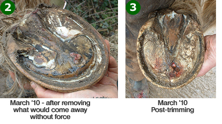
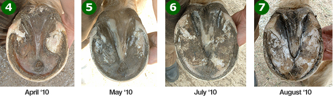 You
can see how his sole continued to recover in these pictures – there is just
over 12 weeks between pictures 2 and 7 The cavity is now (five months later)
little more than a slight depression in his sole. Most importantly, one
month after pictures 2 and 3, he was 100% sound without boots, and crunching
over a rough stony yard to get to his field.
You
can see how his sole continued to recover in these pictures – there is just
over 12 weeks between pictures 2 and 7 The cavity is now (five months later)
little more than a slight depression in his sole. Most importantly, one
month after pictures 2 and 3, he was 100% sound without boots, and crunching
over a rough stony yard to get to his field.
There was obviously veterinary involvement in terms of discussion and antibiotics, and a lot of work by his owner, doing a disinfectant soak, changing dressings and keeping boots/pads and foam in place. Although always a pleasure to deal with, he was not the ideal patient, as he frequently escaped from anywhere we tried to restrict him, shedding dressings and hoof boots on the way, which made his treatment rather tricky.
His front feet are still a work in progress, and I will put more pictures here as his feet improve in shape now that the laminitis is under control.
Case 2
Popeye is a horse I have been trimming since January 2010 at Horseworld Visitor Centre – a rescue, rehabilitation and re-homing charity near Bristol. The pictures are of his right hind hoof. No other management changes were made. He is retired so only does a little in-hand work.
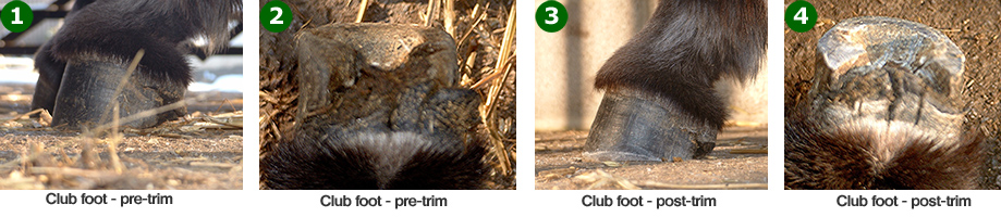
Picture 1: Notice how his lateral heel has collapsed forwards and outwards, the bar has become bent, and there is little surface area at the heels for such a heavy horse (he’s a very feathery cob). The heels are also rather low, and his foot is very asymmetrical.
Pictures 2 – 5 show his lateral heel gradually coming back into a correct place, the bar beginning to straighten out and form a better attachment, with more surface area at his heels. The heels have also gained correct height, and the whole foot is a much better shape.
Case 3
Pixie is a Donkey, also at Horseworld Visitor Centre. She has congenital club foot of her left fore. These photos show how much improvement to foot balance can sometimes be made in one trim …… In pictures 1 and 2 you can see how long her heels are, how far her frog is therefore off of the ground, and how she is beginning to ‘knuckle over’.
Pictures 3 and 4 show her frog just about able to make ground contact, and her heels a much better length – this was done carefully, checking her comfort and tendon length at frequent intervals during trimming.
Other Interesting Pictures
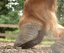
 This
picture (left) shows excessive heel height and growth rings due to
chronic long term laminitis, with some probable acute attacks also involved.
This
picture (left) shows excessive heel height and growth rings due to
chronic long term laminitis, with some probable acute attacks also involved.
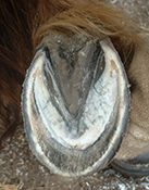
The other picture (right) is taken mid-trim, and nicely shows the huge amount of disruption which can go on in a laminitic foot.
'Molly’ (one of my favourites!) – was about to be put down a
few years ago because she was so dangerous, and is now
a level 3 Parelli horse - she also does general hacking and
riding several times a week – a credit to her owner and rescuer.